Meso Scale Discovery (MSD) increases ELISA linear range by 2-3 orders of magnitude and increases sensitivity by 10-100 times
Percepta's report on top 550 biomedical researchers in North America and Europe shows that MSD (Meso Scale Discovery) based on electrochemiluminescence technology is one of the top 5 immunoassay brands. The advantages of MSD electrochemiluminescence technology and traditional enzyme-linked immunosorbent assay ELISA are as follows:
1. Higher sensitivity and wider linear range
The traditional enzyme-linked immunosorbent assay (ELISA) is based on a microplate reader, and the sensitivity of the microplate reader is often 10-1 pg/ml. MSD electrochemiluminescence technology is mainly based on the super sensitive multi-factor electrochemiluminescence analyzer MESO Sector 600 or Quickplex SQ 120. MSD sensitivity is 0.05pg/ml and the effective linear range is 6 log.
Conventional ELISA has a linear range of 10 pg/ml to 1000 pg/ml, and an Ultra-sensitivity kit of 0.5-10 pg/ml. For a biological experiment, it is often necessary to take into account the samples of the normal control group and the disease group. The concentration of the tested protein in the sample generally ranges from a few pg to several thousand pg, so the linear range of an ELISA kit cannot be balanced at the same time. The detection of high and low abundance proteins requires a lot of pre-experiment for dilution and exploration, which is time consuming and laborious. MSD offers a wide linear range from sub-pic to tens of picograms, effectively placing all samples within the optimal linear range for accurate determination (see image below, red strip for disease sample, yellow) The strip is a normal reference group). At the same time, the sensitivity of MSD can reach 0.05pg/ml. For the research projects in which the protein concentration of the disease is down-regulated in some disease samples, it is more effective to find the difference between the disease group and the normal control group and realize new scientific findings.

2.
Save sample size and achieve multiple detection 
The sample size required for traditional ELISA is generally 50-100 ul. If the expression regulation of 10 cytokines is concerned, it may require as many as 500-1000 ul samples. For many ELISA experiments, the sample size becomes a key issue.
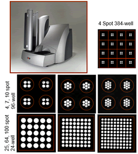
MSD uses dot matrix technology to detect 10 indicators per well in a 96-well graphite electrode. Screening and testing of 100 indicators/holes can also be customized in 24-well plates. Meet different experimental needs. In the sample dosage, the test can be completed with only ≤25 ul of samples, whether single or multiple indicators. More data is available for the study of rare samples.
At present, MSD can quantitatively measure 40 kinds of Biomarker in 100 ul of extremely precious cerebrospinal fluid samples, which is impossible in the prior art.
3. Uniformity, high repeatability
The CV in the MSD plate is <6-8%, <10% between plates, and <15% between batches. At the same time, the MSD kit is valid for up to 30 months, providing the possibility of using the same batch of reagents for long-term research projects.
4. High sample compatibility and small matrix effect
Due to its unique electrochemiluminescence principle, the MSD platform can eliminate many non-specific signals, so it is less affected by the nature of the sample (such as viscous samples, suspended particles, etc.), so for various biological samples. More compatible.
At present, the total sample of samples on the MSD platform mainly includes: serum, plasma, culture supernatant, cell/tissue lysate, cerebrospinal fluid, urine, joint synovial fluid, saliva, sputum, stool extract, eye aqueous humor, various types of irrigation. Basic biological samples such as lotion. For the simplest plasma samples, the MSD is also compatible with plasma samples of different anticoagulants. The difficulty in collecting samples required for the experiment is greatly reduced.
In addition, the matrix effect will affect the accuracy and sensitivity of the experiment. MSD is currently the only immunoassay platform with no matrix effect widely recognized by foreign scientists.
See: Immunol Res; DOI 10.1007/s12026-014-8491-6; T.Maecker
5. The experiment process is simple, fast, and diversified
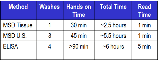
MSD provides a more convenient and faster experimental procedure than ELISA, reducing the manual operation time such as loading to less than 45 minutes, and providing a one-step process of 2.5 hours for rapid detection of high-abundance samples such as culture supernatants. Liberated the labor force and improved the efficiency of scientific research. At the same time, due to the unique electrochemiluminescence technology on the MSD platform, the background signal can be effectively controlled, and the no-clean experimental process can be realized. Please see the MSD process below and the comparison of experimental time:
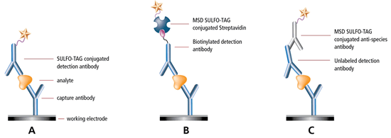
One-step process:
Hypersensitive experimental procedure:
Experimental process time comparison:
6. The signal is stable and unaffected by the color sequential
When ELISA is developed, it is necessary to protect from light to ensure signal stability. At the same time, due to the difference in the timing of the addition of the substrate and the stop solution, data differences due to inconsistencies in the color development time are inevitably generated.
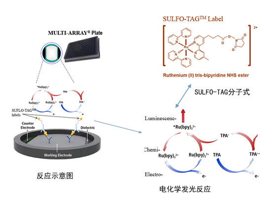
Because MSD is based on electrochemiluminescence technology, the signal molecule SULFO-TAG needs to be generated under the condition of electric excitation. Therefore, the whole experimental process does not need to be protected from light and is not affected by the difference of the technical level of the experimental operators. In addition, no signal is evoked after the experiment completes the addition of all liquids. Only when sent to the instrument, the signal is generated after the electrode is electrically excited, and the excitation of each hole is unified with the signal acquisition time, which avoids the data difference caused by the sequential order of the ELISA substrate and the stop liquid, and the data stability is extremely high. Please see the diagram below for the electrochemiluminescence platform:
7. Open platform, high-load graphite electrode, diversified experimental scheme, and more savings in coating dosage.
The graphite electrode plate used in the MSD platform is a polymer material with a 3D structure on the surface and inside, which can increase the effective load to 10-100 times that of the conventional ELISA plate. Therefore, proteins, small peptides, polysaccharides, nucleic acids, virus particles, cell membranes, living cells, etc. can be immobilized at the bottom of the plate to realize different experimental detection schemes.
3D structure diagram of the bottom of the board
Diversified experimental protocol
At the same time, due to the different loading and surface hydrophobicity modification, the MSD plate can use the traditional method Solution Coat in the coating scheme, or the Dot Coat method, and coat 2-5ul protein spots in the center of the plate. . Protein usage requires only 1/10-1/5 of Solution Coat, and the coating time takes only one hour, saving time and effort.
8 . The technology is mature and reliable. European and American researchers have started using the "Meso Scale Discovery" replacement / upgrade ELISA 10 years ago .
9. Some of the test indicators are as follows:
Multi-factor detection combination | Detection Indicator |
Human Biomarker 40-Plex Kit (1 Plate) V-PLEXTM | bFGF, CRP, Eotaxin, Eotaxin-3, Flt-1, GM-CSF, ICAM-1, IFN-γ, IL-1α, IL-1β, IL-10, IL-12 p70, IL-12/IL-23p40 , IL-13, IL-15, IL-16, IL-17A, IL-2, IL-4, IL-5, IL-6, IL-7, IL-8, IL-8 (HA), IP- 10, MCP-1, MCP-4, MDC, MIP-1α, MIP-1β, PlGF, SAA, TARC, Tie-2, TNF-α, TNF-β, VCAM-1, VEGF, VEGF-C, VEGF- D |
Neuroinflammation Panel 1 (hu) Kit (1 Plate) V-PLEXTM | bFGF, CRP, Eotaxin, Eotaxin-3, Flt-1, ICAM-1, IFN-γ, IL-1α, IL-1β, IL-10, IL-12/IL-23p40, IL-13, IL-15, IL-16, IL-17A, IL-2, IL-4, IL-5, IL-6, IL-7, IL-8, IP-10, MCP-1, MCP-4, MDC, MIP-1α, MIP-1β, PlGF, SAA, TARC, Tie-2, TNF-α, TNF-β, VCAM-1, VEGF, VEGF-C, VEGF-D |
Human Cytokine 30-Plex Kit (1 Plate) V-PLEXTM | Eotaxin, Eotaxin-3, GM-CSF, IFN-γ, IL-1α, IL-1β, IL-10, IL-12 p70, IL-12/IL-23p40, IL-13, IL-15, IL-16 , IL-17A, IL-2, IL-4, IL-5, IL-6, IL-7, IL-8, IL-8 (HA), IP-10, MCP-1, MCP-4, MDC, MIP-1α, MIP-1β, TARC, TNF-α, TNF-β, VEGF |
NHP Cytokine 24-Plex Kit (1 Plate) V-PLEXTM | Eotaxin-3, GM-CSF, IFN-γ, IL-1β, IL-10, IL-12/IL-23p40, IL-15, IL-16, IL-17A, IL-2, IL-5, IL- 6, IL-7, IL-8, IL-8 (HA), IP-10, MCP-1, MCP-4, MDC, MIP-1α, MIP-1β, TARC, TNF-β, VEGF |
Proinflammatory Panel 1 (rat) Kit (1 Plate) V-PLEXTM | IFN-γ, IL-1β, IL-10, IL-13, IL-2, IL-4, IL-5, IL-6, KC/GRO, TNF-α |
Chemokine Panel 1 (human) Kit (1 Plate) V-PLEXTM | Eotaxin, Eotaxin-3, IL-8 (HA), IP-10, MCP-1, MCP-4, MDC, MIP-1α, MIP-1β, TARC |
Proinflammatory Panel1 (mouse) Kit (1 Plate) V-PLEXTM | IFN-γ, IL-1β, IL-10, IL-12 p70, IL-2, IL-4, IL-5, IL-6, KC/GRO, TNF-α |
Proinflammatory Panel1 (human) Kit (1 Plate) V-PLEXTM | IFN-γ, IL-1β, IL-10, IL-12 p70, IL-13, IL-2, IL-4, IL-6, IL-8, TNF-α |
Cytokine Panel 1 (human) Kit (1 Plate) V-PLEXTM | GM-CSF, IL-1α, IL-12/IL-23p40, IL-15, IL-16, IL-17A, IL-5, IL-7, TNF-β, VEGF |
Human Proinflammatory Panel I (4-Plex) (1 Plate) V-PLEXTM | IFN-γ, IL-1β, IL-6, TNF-α |
Human Proinflammatory Panel II (4-Plex) (1 Plate) V-PLEXTM | IL-1β, IL-6, IL-8, TNF-α |
Chemokine Panel 1 (NHP) Kit (1 Plate) V-PLEXTM | Eotaxin-3, IL-8 (HA), IP-10, MCP-1, MCP-4, MDC, MIP-1α, MIP-1β, TARC |
Chemokine Panel 1 (NHP) Kit (1 Plate) V-PLEXTM Plus | Eotaxin-3, IL-8 (HA), IP-10, MCP-1, MCP-4, MDC, MIP-1α, MIP-1β, TARC |
Proinflammatory Panel 1 (NHP) Kit (1 Plate) V-PLEXTM | IFN-γ, IL-1β, IL-10, IL-2, IL-6, IL-8 |
Cytokine Panel 1 (NHP) Kit (1 Plate) V-PLEXTM | GM-CSF, IL-12/IL-23p40, IL-15, IL-16, IL-17A, IL-5, IL-7, TNF-β, VEGF |
Angiogenesis Panel 1 (human) Kit (1 Plate) V-PLEXTM | bFGF, Flt-1, PlGF, Tie-2, VEGF, VEGF-C, VEGF-D |
Vascular Injury Panel 2(human) Kit (1 Plate) V-PLEXTM | CRP, ICAM-1, SAA, VCAM-1 |
Ab Peptide Panel 1 (4G8) Kit (1 Plate) V-PLEXTM | Abeta 38, Abeta 40, Abeta 42 |
Ab Peptide Panel 1 (6E10) Kit (1 Plate) V-PLEXTM | Abeta 38, Abeta 40, Abeta 42 |
LBD Tengquan Headquarters
Room 211, Building 1, 1133 Halle Road, Zhangjiang Hi-Tech Park, Shanghai
Phone: 086-21-51088608
Fax: 086-21-50478211
mailbox:
Organic Ginseng Extract
Organic red ginseng extract is extracted from dried red ginseng roots. The raw material of red ginseng is fresh ginseng. After fresh ginseng is steamed, its skin turns red, so it is called red ginseng.
During the red ginseng cooking process, due to the heat treatment, a chemical reaction occurs, and the composition changes. It will produce new ingredients that are not found in water ginseng and white ginseng, namely, red ginseng`s unique physiologically active substance G-Rh2, ginsengtriol, maltol, and so on. G-Rh2 and ginsenotriol can inhibit the growth of cancer cells, while maltol has an antioxidant effect.
Ginseng Extract,Organic Red Ginseng Extract,Ginseng Planting Base,Dried Red Ginseng Roots
Organicway (xi'an) Food Ingredients Inc. , https://www.organic-powders.com


