Overview: April 20th Micro-CT angiography was performed on mice inoculated with liver cancer cells at the Sun Yat-sen University Cancer Hospital to observe the preparation of tumor models. Experimental results show that Branch Kai Sheng ZKKS-MCT-III type micro-CT system is clear in vivo, non-invasive, continuous dynamically distributed tumors observed in mice, the growth percentage of volume change of tumor size and In the case, the experimental results have achieved the intended purpose.
Experiment date : 2010.04.20
OBJECTIVE : To detect the tumor model preparation and tumor growth of C57 mice inoculated with liver tumor cells in situ; to use common Kunming mice to try the contrast effect of common contrast agents on organs.
Experimental instruments and reagents :
ZKKS-MCT-III micro-CT system, ZKKS-MCT-III micro-CT system software;
Contrast agent: Fenestra VC (ART Advanced Research Technologies Inc., QC, Canada)
Clinical use of iohexol (GE, Omnipaque 350 mg I/ml);
Consumables such as syringes necessary for the experiment.
Experimental object :
Table 1 Mouse condition for experiment
Mouse name | Weight ( g ) | Modeling situation | Inoculation cycle |
C57 is numbered M | 21.0 | Liver in situ vaccination | 4-5 weeks |
C57 number is N | 19.6 | Liver in situ vaccination | 4-5 weeks |
KM number is P | 22.0 | Null | Null |
Laboratory personnel : Liang Yi, Liu Junting, Liu Yanqun, Yan Maojie, Chen Xiaofeng
Experimental steps :
1. M mice ( C57 mice 21.0 g )
(1) Preparation before imaging
11:02 M mice were anesthetized;
11:08 tail vein injection of small animal contrast agent Fenestra VC
(ART Advanced Research Technologies Inc., QC, Canada)
(2) micro-CT data acquisition
14:38 Start scanning, the parameter settings are as follows:
X-ray tube voltage: 50KVp; power: 60W;
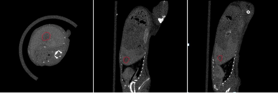
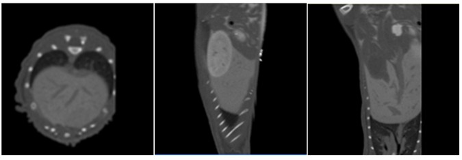
The results of the Micro-CT scan are shown in Figure 1.
Figure 1 Cross-sectional, sagittal, coronal view of C57 mice numbered M
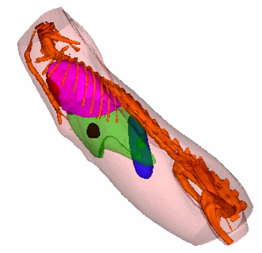
Figure 2 Three-dimensional display of tumor tissue obtained by micro-CT reconstruction and segmentation of the slice
(The brown tissue in the figure is tumor tissue, red is heart tissue, purple is lung,
Green is liver, blue is spleen, golden yellow is bone, meat red is fat and muscle)
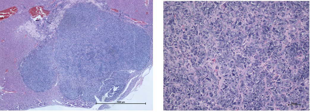
Figure 3 Histopathological section of mouse liver tumor
(The left side is a 4x microscopic observation, and the right side is a 20x microscopic observation.)
The mice were collected at 11:36, 12:22, 12:38, and 14:38 under the same parameters. The data showed similar results. It was obvious that the diameter was about 3 mm from the slice. The approximately spherical tumor tissue was measured by a software volume of 13.0107 mm 3 .
On the third day after the end of the experiment, Dr. Liang Yi from the Zhongshan Cancer Hospital did not find any visible tumor masses after dissection. After pathological biopsy, he found tumor lesions in the liver (see Figure 3). The results of the biopsy were identical to the CT measurements. .
Experiment (1) Summary : Fenestra VC small animal contrast agent was successfully injected in the experiment. The biggest advantage of this contrast agent is that it can maintain long-term contrast effect in the body. The experimental results fully satisfied the purpose of monitoring the preparation of the model, and found a tumor with a diameter of about 3 mm and a total volume of 13.0107 mm 3 .
2. N mice ( C57 mice 19.6 g )
(1) Preparation before imaging
11:46 Anesthetize the mice numbered N;
12:20 Intravenous injection of Fenestra VC
(ART Advanced Research Technologies Inc., QC, Canada)
(2) micro-CT data acquisition
14:15 Start scanning, the parameter settings are as follows:
X-ray tube voltage: 50KVp; power: 60W;
The results of the Micro-CT scan are shown in Figure 1.
The mice were found dead after the end of the scan.
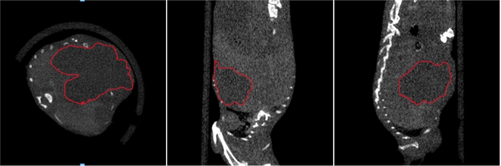
Figure 4 is a cross-sectional, sagittal, and coronal view of C57 mice numbered N
(where the area indicated in the red line is tumor tissue)
Figure 5: Three-dimensional display of tumor tissue obtained by micro-CT reconstruction and segmentation of the slice
(The golden color is the bone in the picture, the tumor is the tumor in the brown, and the transparent tissue in the meat red is the adipose tissue.)
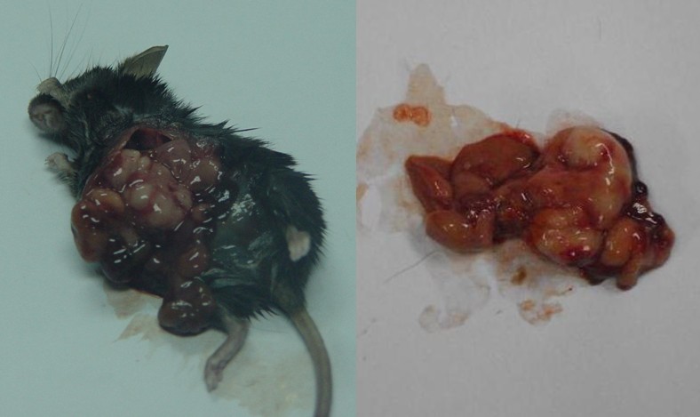
To further confirm that the micro-CT scan detected the tumor tissue, we dissected the mouse and dissected the tumor tissue to find that the tumor had formed a large mass after about 5 weeks of growth, as shown in Figure 6. . The volume occupied by the tumor can be calculated by the micro-CT system software to be 1.0631cm 3 .
Figure 6: The tumor tissue was found after dissection of C57 mice numbered N.
(The left picture shows the C57 number N mouse after opening the chest, and the right picture shows the liver and white tumor mass tissue)
Experiment (2) Summary : Fenestra VC small animal contrast agent was successfully injected in the experiment. The mice died 2 hours after the contrast injection, and the contrast agent was not fully metabolized in the mouse organs. The cause of death analysis is that the tumor mice are too weak. In addition, reconstruction with a size slightly larger than the width of the mouse during micro-CT reconstruction will make the resolution of the image clearer.
3. P mice (Kunming mice 22 g )
To further validate the contrast effect of clinical iodine (GE, Omnipaque 350 mg I/ml) on the contrast of mouse organ tissue, we combined the experience of previous iohexol contrast agent to summarize the common Kunming mice. Contrast imaging of the organ was performed.
Experimental steps:
(1) Preparation before imaging
Pre-imaging the experimental mice on April 21
Mice were anesthetized at 14:30 on April 22.
Scanning mice starting at 15:00 on April 22
X-ray tube voltage: 55KVp; power: 60W;
The results of the Micro-CT scan are shown in Figure 5.
Figure 7 Contrast results of visceral tissue using clinical iohexol contrast agent
Experiment (3) Summary : The clinical use of iohexol contrast agent can also be used for angiography of organs. This method of angiography can greatly save the cost of reagents for small animal contrast agents. In the follow-up tumor research, this low-cost iohexol contrast agent is recommended, and a better contrast effect can be achieved.
Conclusion: The ZKKS-MCT-III micro-CT system can be used to observe the growth and volume changes of tumor cells in mice in vivo, non-invasively and continuously. The experimental data show that not only can the instrument be used to monitor the modeling, but also the micro-CT system can accurately observe the tumor volume and shape during the treatment. . This experiment successfully achieved the expected results.
Oxygen Cylinder Filling System
With PSA principles, ETR Oxygen Cylinder Filling system can produce 93%±3% purity oxygen gas from compressed air directly. ETR oxygen cylinder filling system is consisted of Atlas screw air compressor, refrigerated air dryer, compressed air filter, air buffer tank, ETR Oxygen Plant, oxygen buffer tank, oxygen booster, cylinder filling station and HMI control cabinet.
Compressed air is purified through the air dryer and filters to a certain level for main plant to work with. Air buffer is incorporated for smooth supply of compressed air thus to reduce fluctuation of compressed air source. The plant produces oxygen with PSA (pressure swing adsorption) technology, which is a time proven oxygen generation method. Oxygen of desired purity at 93%±3% is delivered to oxygen buffer tank for smooth supply of product gas. Oxygen in buffer tank is maintained at 4bar pressure. With an oxygen booster, the oxygen pressure can be to 150bar and fill in cylinders.

Onsite Oxygen Cylinder Filling System
Onsite Oxygen Cylinder Filling System,Oxygen Refilling System ,Oxygen Cylinder Filling,Oxygen Refilling Machine
Hunan Eter Medical Co., Ltd. , https://www.eter-tech.com